MRI Services

Brain

Because the MRI scan gives very detailed pictures it is the best technique when it comes to finding tumours (benign or malignant abnormal growths) in the brain. If a tumour is present the scan can also be used to find out if it has spread into nearby brain tissue. The technique also allows us to focus on other details in the brain. For example, it makes it possible to see the strands of abnormal tissue that occur if someone has multiple sclerosis and it is possible to see changes occurring when there is bleeding in the brain, or find out if the brain tissue has suffered lack of oxygen after a stroke. Be able to show a clear image of cerebral vessels.

    MRI Brain scan is needed for a person who has a neurological sign:
  • Chronic headache
  • Stroke
  • Brain tumor
  • Abnormal vessels in the brain
  • Vertigo
  • Convulsion

Abdomen

Magnetic resonance imaging (MRI) is a diagnostic technique that uses a cylindrical magnet and radio waves to produce cross-sectional images of organs and structures within the body, with superior contrast resolution. Additional advantages of MRI include direct multi-planar imaging capability, without ionizing radiation exposure. The body’s hydrogen atoms react to the magnetic field and pulses of radio waves and differences in the detected signal intensities are displayed on a computer-generated image.

MRI Upper Abdomen provides extraordinary detail of the images inside the Abdomen . The difference between normal and abnormal tissue is often clearer on the MRI scan than on the CT scan. Be used for examining the abdominal parts (such as liver, kidney, pancreas and spleen) . MRI can be used for problem-solving evaluation of ambiguous or equivocal findings on CT, Ultrasound or other diagnostic examinations.

Magnetic Resonance Imaging (MRI) is considered medically necessary for the evaluation of the following abdominal indications:

  • Adrenal masses including differentiation of adenoma from metastasis.
  • Focal liver lesions (e.g., cysts, metastases, hemangiomas).
  • Conditions for which a CT scan is indicated but who have a contraindication to the use of CT.
  • Non-cystic or indeterminate renal parenchymal mass.
  • Define extension of a tumor into the renal vein and inferior vena cava.
  • To define the extrarenal extent of renal neoplasm.
  • Staging of malignancies.
  • Abnormalities noted on other imaging studies, which require additional clarification.
  • Additional information regarding timing MRI of the abdomen:
  • MRI may provide more sensitive imaging if US and/or CT are inconclusive.
  • MRI imaging is more sensitive for suspected cavernous hemangioma, hemachromatosis, hemosiderosis, and fatty liver.
  • The MRI is preferred for liver lesion characterization when there is existing liver disease (hepatitis, cirrhosis).

MRI scan is needed for a person who has a symptom:

  • Difficulty in swallowing
  • Jaundice
  • Abdominal pain
  • Ascites(Abnormal accumulation of fluid in the cavity of the abdominal)
  • Viral Hepatitis B carrier

Pelvis

The pelvic area contains the reproductive organs.

  • In women, it includes the womb (uterus), cervix, ovaries, and fallopian tubes.
  • In men, it includes the prostate gland and testicles.

For women, pelvic MRI provides a detailed look at the ovaries and uterus and is often used to follow up an abnormality seen on ultrasound. It is also used to evaluate the spread of cancer of the uterus.

For men, pelvic MRI is sometimes used to check those diagnosed with prostate cancer. The primary indication for MRI of the prostate is the evaluation of prostate cancer. The test is commonly used after a prostate biopsy has confirmed cancer in order to determine if the cancer is confined to the prostate, or if it has spread outside the walls of the prostate gland.

Occasionally, MRI of the prostate is used to evaluate other prostate problems, including : infection (prostatitis) or prostate abscess , an enlarged prostate, called benign prostatic hyperplasia (BPH). Pelvic MRI is also used to look at the bones and muscles of the pelvis.

MRI Pelvis may be done if a female has any of the following signs or symptoms:

  • Abnormal vaginal bleeding
  • A mass in the pelvis (felt during a pelvic exam or seenon another imaging test)
  • A pelvic mass that occurs during pregnancy
  • Endometriosis (usually only done after ultrasound)
  • Lower abdominal pain
  • Unexplained infertility (usually only done after ultrasound)
  • Unexplained pelvic pain (usually only done after ultrasound)

MRI Pelvis may be done if a male has any of the following signs or symptoms:

  • Lumps or swelling in the testicles or scrotum
  • Undescended testicle (unable to be seen using ultrasound)
  • Unexplained pelvic or lower abdominal pain
  • Unexplained urination problems, including trouble starting or stopping urinating

A pelvic MRI may be done in both males and females who have:

  • Abnormal findings on an x-ray of the pelvis
  • Birth defects of the hips
  • Injury or trauma to the hip area
  • Unexplained hip pain

A pelvic MRI is also frequently done to see if certain cancers have spread to other areas of the body. This is called staging. Staging helps guide future treatment and follow-up and gives you some idea of what to expect in the future. A pelvic MRI may be used to help stage cervical, uterine, bladder, rectal, prostate and testicular cancers.

Results depend the nature of the problem. Different types of tissues send back different MRI signals. For example, healthy tissue sends back a slightly different signal than cancerous tissue.

Abnormal results in a woman may be due to:

  • Adenomyosis
  • Bladder cancer
  • Cancer of the renal pelvis or ureters that has spread from the kidneys
  • Cervical cancer
  • Colorectal cancer
  • Congenital defect of the reproductive organs
  • Endometrial cancer
  • Endometriosis
  • Ovarian cancer
  • Ovarian growths
  • Problem with the structure of the reproductive organs, such as the fallopian tubes
  • Uterine fibroids

Abnormal results in a man may be due to:

  • Bladder cancer
  • Cancer of the renal pelvis or ureters that has spread from the kidneys
  • Colorectal cancer
  • Prostate cancer
  • Testicular cancer

Abnormal results in both males and females may be due to:

  • Avascular necrosis
  • Birth defects of the hip joint
  • Bone tumor
  • Hip fracture
  • Osteoarthritis
  • Osteomyelitis

Breast

MRI of the breast is a tool for detecting and staging breast cancer and other breast abnormalities. MRI of the breast offers valuable information about many breast conditions that cannot be obtained by other imaging modalities, such as mammography or ultrasound.

    Without contrast material, an MRI of the breast can show:
  1. breast tissue density.
  2. cysts.
  3. enlarged ducts.
  4. hematomas.
  5. leaking or ruptured breast implants.
  6. the presence of enlarged lymph nodes.
    By comparing breast images taken before and after contrast material injection, an MRI exam can determine:
  1. if there are breast abnormalities.
  2. whether an abnormality looks benign (non-cancerous) or malignant (cancerous).
  3. the size and location of any abnormality that looks malignant.
    MR imaging of the breast is performed to:
  1. assess multiple tumor locations, especially prior to breast conservation surgery.
  2. identify early breast cancer not detected through other means, especially in women with dense breast tissue and those at high risk for the disease.
  3. evaluate abnormalities detected by mammography or ultrasound.
  4. distinguish between scar tissue and recurrent tumors.
  5. determine whether cancer detected by mammography, ultrasound, or after surgical biopsy has spread further in the breast or into the chest wall.
  6. assess the effect of chemotherapy.
  7. provide additional information on a diseased breast to make treatment decisions.
  8. determine the integrity of breast implants.

How is the procedure performed?

MRI examinations may be performed on outpatients . You will be positioned on the moveable examination table. Straps and bolsters may be used to help you stay still and maintain the correct position during imaging. For an MRI of the breast, you will lie face down on your stomach with your breasts hanging freely into cushioned openings, which are surrounded by a breast coil, which is a signal receiver that works with the MRI unit to create the images. If a contrast material will be used in the MRI exam, a nurse or technologist will insert an intravenous (IV) line into a vein in your hand or arm. A saline solution will drip through the IV to prevent blockage of the IV line until the contrast material is injected.

Joints

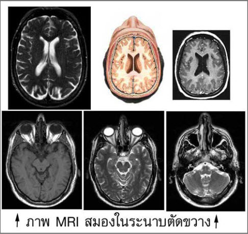
Because MRI can give such clear pictures of soft tissue structures near and around bones.

    • MRI is usually the best choice for examining the:


  • body’s major joints.
  • soft tissues of the extremities (muscles and bones).

MR imaging is typically performed to diagnose or evaluate:

  • degenerative joint disorders such as arthritis and meniscus tears (knee) or labral tears (shoulder and hip).
  • locate and identify the cause of pain,swelling or bleeding in the tissues in and around the joints and bones.
  • allow the physician to clearly see even very small tears and injuries to tendons, ligaments and muscles that cannot be seen on xrays.
  • fractures (in selected patients).
  • deterioration of joint surfaces , joint abnormalities.
  • sports-related injuries and work-related disorders caused by repeated strain, vibration or forceful impact.
  • infections (such as osteomyelitis).
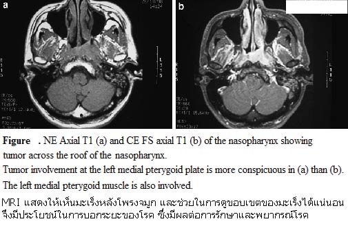
  • tumors (primary tumors and metastases) involving bones and joints.

INDICATIONS AND SPECIFIC TECHNIQUES FOR MRI OF THE MUSCULOSKELETAL SYSTEM

Soft Tissues

Spectroscopic fat saturation or short inversion time inversion recovery (STIR) fat suppressed images may be helpful in evaluating soft tissue masses and subtle bone injuries.

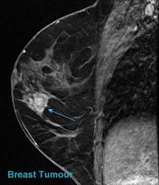
MRI is indicated for the evaluation of traumatic muscle and tendon injuries, hematomas, compartment syndromes , entrapment syndromes, tendinosis, tenosynovitis, and bursitis. MRI is useful for the evaluation of infections, abscesses and myositis. It is indicated for theevaluation of masses such as simple non-neoplastic cysts, abscesses, ganglion cysts, parameniscal cysts, hematomas, muscle tears, and ligament and tendon tears. It is also useful for the detection, staging, and characterization of benign and malignant soft tissue neoplasms and for the follow-up evaluation of neoplastic disease after therapy. (Intravenous contrast agents may be helpful in evaluating potentially malignant disease . For the evaluation of genetic muscle diseases, MR spectroscopy may also be helpful.

Joints

1. Diseases affecting all joints

MRI is indicated for the evaluation of traumatic injuries to joints and adjacent muscles, tendons, and ligaments , selected articular cartilage injuries , bursitis and synovitis from overuse, fragment stability and cartilage status in osteochondritis dissecans , posttraumatic osteonecrosis and degenerative joint disease , loose bodies, and tenosynovitis. MRI is useful in the evaluation of joint infections, noninfectious inflammatory joint disease such as rheumatoid and the seronegative arthritides , overuse synovitis, and tenosynovitis. It is useful for the evaluation of ganglion cysts , bursal cysts with bursitis , abscesses, benign neoplastic masses, and primary and metastatic masses . MRI is indicated for the evaluation of osteonecrosis including avascular necrosis , and degenerative joint disease .

2. Diseases affecting specific joints
  • Shoulder

    Imaging planes should be selected to coincide with the primary axes of the shoulder. Long TR, double echo coronal images should be obliqued electronically parallel to the supraspinatus muscle or perpendicular to the glenoid fossa of the scapula. (T1W) or (T2W) sagittal images should be obtained perpendicular to the oblique coronal images. Turbo or fast spin echo images are often utilized commonly with fat suppression in the oblique coronal plane. The field-ofview (FOV) should be 12-16 cm. The slice thickness should be less than 5 mm and spatial resolution less than 1 mm. Images in the oblique coronal plane should be acquired with long TR and short and long TE spin echoes such as with a TR of 2,000 msec and TEs of 20 and 80 msec (51). Images in the oblique sagittal plane require contrast adequate for anatomical evaluation, such as TR 500, TE 15 msec or TR 2,000 msec and TEs of 20 and 80 msec. The axial images require good anatomical information as well as images sensitive for fluid. Gradient echo or long TR double spin echo images are adequate in this plane. Acquisition matrices of 192 x 256 are desirable with the number of excitations/averages selected to ensure adequate signal-tonoise.

    MRI is indicated for the evaluation of shoulder pain of undetermined etiology, detection and staging of rotator cuff degeneration (tendinosis) and tears, impingement syndromes , labral degenerative changes and tears, biceps tendon disease and dislocation, suprascapular notch syndrome, glenohumeral ligament injuries, coracoclavicular and acromioclavicular separations, subacromial bursitis, and joint anatomy after bony fracture. Suspected instability and labral tears may require intraarticular contrast administration for optimal detection (either MRI after gadolinium chelate or saline injection, or CT arthrography).

  • Elbow

    Local surface coils and FOV of less than 12 cm are recommended.

    MRI is indicated for the evaluation of medial epicondylitis (tennis elbow), fractures in children, osteochondral defects, and osteonecrosis.

  • Wrist, Hand, and Fingers

    Local surface coils and FOV of 10 cm or less are recommended. Imaging planes should be carefully selected to be in the true coronal, sagittal, and axial planes. Electronic angulation may be necessary to insure proper plane selections. An MR-compatible brace on the wrist may be useful to ensure uniform wrist positioning.

    MRI is indicated for the detection and evaluation of the cause of carpal tunnel syndrome, tendon and ligamentous injuries, triangular fibrocartilage injuries, extensor and flexor tenosynovitis, de Quervain’s syndrome , Kienbach’s disease , injuries of the flexor and extensor tendons , tenosynovitis, and masses.

  • Hip

    Most imaging of the hips is performed without surface coils because of the difficulties of surface coil design for hip anatomy. T1W coronal images are sensitive for osteonecrosis and fractures. T2W images or high-resolution three-dimensional gradient echo images may be useful for evaluating fine anatomical details, especially in children. Subtle bone injuries are common and clinically relevant and may require fat suppressed coronal images for optimal sensitivity.

    MRI is indicated for the evaluation of avascular necrosis , congenital hip dislocation, congenital hip dysplasia, transient osteoporosis, and iliopsoas bursitis.
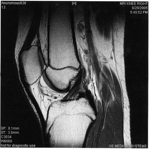
  • Knee

    Imaging should be performed in at least two planes utilizing at least one (T2W) technique. FOV should be less than 16- cm, pixel dimension less than 1 mm, and slice thickness less than 5-mm. Acquisition matrices of 192 x 256 are advantageous. Magnified, narrow window width display of short TE images containing the menisci may be of benefit in interpretation of meniscal disease.

    MRI is indicated for the evaluation of knee pain of undetermined etiology, meniscal tears, discoid menisci, suspected cruciate and collateral ligament tears, bone contusions (trabecular fractures), patellar chondromalacia, patellar tracking abnormalities, popliteal cysts and aneurysmsec, pes anserinus bursitis, prepatellar bursitis, pigmented villonodular synovitis, evaluation of anterior cruciate ligament reconstructions , and meniscal and ganglion cysts.

  • Ankle and Foot

    FOV less than 12 cm are recommended with appropriately designed local coils. Images should be obtained in the coronal, sagittal, and axial planes with respect to the anatomic region of interest. Suspected disease in the tendons coursing through the ankle may be best evaluated with angled axial images, perpendicular to the tendon at the level of suspected disease. Long TR double echo images and STIR images are advantageous in evaluating tendon and subtle bony injuries. When a tear of the Achilles tendon is suspected, a large FOV sagittal image may be warranted to evaluate the extent of possible retraction of the superior segment. When subtle findings are present, imaging of the contralateral ankle may be helpful.

    MRI is indicated for the evaluation of ankle pain of undetermined etiology , tendon and ligament injuries , sinus tarsi syndrome , tarsal tunnel syndrome , plantar fasciitis, masses , postoperative evaluation of Achilles tendon repair , diabetic foot disease , and neurogenic joint disease.

  • Temporomandibular Joint (TMJ)

    Local surface coils are necessary with FOVs of less than 12-cm. Slice thickness should be 3 mm or less and spatial resolution should be less than 1 mm. Sagittal highresolution (T1W) images are usually performed to determine disc position. If inflammatory disease is present, then long TR, long TE (T2W) images are helpful in evaluating pathology . Kinematic imaging with rapid T1 or gradient echo images may be helpful in characterizing disc placement abnormalities . Since TMJ disease may be bilateral in up to 80% of affected patients, both joints should be imaged, ideally simultaneously .

    MRI is indicated for the evaluation of joint pain and clicking , abnormal disc displacement , degenerative joint disease , osteonecrosis of the mandibular condyle , foreign body reaction , inflammatory joint disease , trauma , ganglion cysts , and synovial chondromatosis .

Spines

An MRI of the spine shows the anatomy of the vertebrae that make up the spine,as well as the disks, spinal cord and the spaces between the vertebrae through which nerves pass. Depending on the location of symptoms,only part of the spine may be imaged: the cervical (neck) portion, the thoracic (chest) spine or the lumbar (lower) spine.

MR imaging is performed to:

  • Assess the spinal anatomy.
  • visualize anatomical variations and diseased tissue in the spine.
  • help plan surgeries on the spine such as decompression of a pinched nerve or spinal fusion.
  • monitor changes in the spine after an operation, such as scarring or infection.
  • guide the injection of steroids to relieve spinal pain.
  • assess the disks – bulging, degenerated or herniated intervertebral disk : a frequent cause of severe lower back pain and sciatica.
  • evaluate compressed (or pinched) and inflamed nerves.
  • explore possible causes in patients with back pain (compression fracture for example).
  • image spinal infection or tumors that arise in, or have spread to, the spine.
  • assess children with daytime wetting and an inability to fully empty the bladder.

Spine MRI may reveal disorders such as:

 
  • Cervical disk disorders
  • Degenerative lesions of the spinal cord
  • Enlarged lymph nodes near the spine
  • Herniated disk
  • Lumbar disk disorders
  • Spinal cord compression
  • Syringomyelia
  • Tumors of the spine
  • Multiple sclerosis
  • Myelomeningocele (children)

Benefits vs. risks of MRI of the spine

Benefits
  • MRI is a noninvasive imaging technique that does not involve exposure to radiation.
  • MR images of the spine are clearer and more detailed than with other imaging methods. This detail makes MRI an invaluable tool in early diagnosis and evaluation of many spinal conditions, including tumors.
  • MRI has proven valuable in diagnosing a broad range of conditions, including but not limited to congenital conditions, chronic spinal cord diseases (such as multiple sclerosis), bone abnormalities (e.g., fracture) , disk conditions (e.g., herniated disk), vascular anomalies and tumors.
  • MRI enables the detection of abnormalities that might be obscured by bone with other imaging methods.
  • The contrast material used in MRI exams is less likely to produce an allergic reaction than the iodine-based materials used for conventional x-rays and CT scanning.
  • MRI demonstrates abnormalities, injuries and diseases in the spinal region that may not be visualized with other imaging methods.
  • MRI is very useful for evaluating spinal injuries It is especially helpful for diagnosing or ruling out acute compression of the spinal cord when clinical examination shows muscle weakness or paralysis.
  • MRI is able to detect subtle changes in the vertebral column that may be an early stage of infection or tumor. The procedure may be better than CT scanning for evaluating tumors, abscesses and other masses near the spinal cord.
Risks
  • The MRI examination poses almost no risk to the average patient when appropriate safety guidelines are followed. If sedation is used there are risks of excessive sedation. The technologist or nurse monitors your vital signs to minimize this risk.
  • Although the strong magnetic field is not harmful in itself, medical devices that contain metal may malfunction or cause problems during an MRI exam.
  • There is a very slight risk of an allergic reaction if contrast material is injected. Such reactions usually are mild and easily controlled by medication.
  • Nephrogenic systemic fibrosis is currently a recognized, but rare, complication of MRI believed to be caused by the injection of high doses of MRI contrast material in patients with poor kidney function.

Whole body

You cannot deny that any disease can undesirably visit us some day. 
What can guarantee that we are healthy? 

Symptoms of illness can indicate body abnormality so that we can perceive them and go to see a physician in a timely manner. 
However, some illnesses occur internally and they do not show any symptoms, so it may be too late when we realize them. 

This is a new era of medicine using a high technology equipment transmitted to create an image on the screen of the various parts of the body. 
MRI (Magnetic Resonance Imaging) is considered ideal modality for the screening that has both high sensitivity and specificity without radiation hazard. 

MRI whole-body scan checkup program can find a lesion in an asymptomatic patient provides more treatment options, offers a better prognosis and cuts down on expense compared to when lesions are found in later stages.

Head & neck

MRI is very useful in the head and neck area, particularly for its ability to discriminate variations in soft tissue. 
However, it will not delineate bony erosion and a CT may be necessary for evaluation for bone adjacent to a lesion. A CT is also more advantageous in imaging the paranasal sinus,as any inflammation of mucosa or thickened secretions will appear as an area of high signal intensity on MRI. 
MIR can also be used to evaluate blood flow. Flow can be detected and mapped by MR and magnetic resonance angiography is, in many cases, replacing traditional angiography. 
In general, MRI is most often utilized as the primary imaging modality when evaluating tumor spread in the paranasal sinuses, cavernous sinuses, dura, brain, nasopharynx, oropharynx, palate, base of tongue, and floor of mouth. That is, the closer to the skull base, MRI offers more advantages over CT. 
MRI is also the modality of choice for evaluating perineural tumor spread. 
The detection of retropharyngeal adenopathy also remains almost solely in the realm of CT and MRI and the presence of such nodal metastasis requires modification of both surgical and radiation fields. 
It is also estimated that about 5% of clinically silent metastatic nodes will be identified on CT and MRI and thus the most thorough assessment of the neck is by combined clinical and imaging evaluation. Imaging also best demonstrates extranodal (extracapsular) tumor extension and if that disease has involved the great vessels or bone. 
MRI would be better than CT if cavernous sinus thrombosis is considered.

Lesions are well visualized with MRI :

  • Tongue
  • Oropharynx
  • Nasopharynx
  • Trachea
  • Thyroid
  • Parathyroid
  • Salivary glands
  • Abnormalities of the throat,
  • Inner ear
  • Eyes
  • Temporomandibular joint
  • Cerebellar pontine angle

MRI has been used for the neoplastic diseases for:

  • Diagnosis
  • Staging
  • Pre-operative evaluation
  • Evaluation for response to treatment
  • Follow-up
  • Restaging
  • Treatment planning for radiation therapy
  • Treatment planning for palliation therapy

MRI of the Head and Neck is considered medically necessary when history or current clinical status meets any of the following criteria:

  • Undiagnosed mass on exam or other imaging studies.
  • Congenital Anomaly requiring treatment.
  • Suspected Wegner’s Granulomatosis.
  • Anosmia.
  • Recurrent epistaxis.
  • Abnormalities noted on other imaging or endoscopic studies which require additional clarification.
  • MRI allows an estimate of tumor spread into surrounding soft-tissue areas, such as the anterior cranial fossa and the retro maxillary space.
  • The MRI is indicated for sudden or progressive proptosis and sudden vision loss in the adult.
  • Known malignancy for diagnosis, staging or evaluation for response to treatment or pre-operative evaluation.
  • Planned treatment for radiation therapy.
  • Unexplained Hearing loss.
  • Significant tinnitus or vertigo.
  • Suspected acoustic neuroma or other tumor or preoperative treatment of acoustic neuroma or other tumor.
  • Significant infection unresponsive to medical treatment, suspected abscess, mastoiditis.
  • Cholesteotoma.
  • MRI better for sensirineural hearing loss.
  • Some indications may require studies of the temporal bone with specific attention to internal auditory canal.
  • MRI is preferred for evaluation of fibrous ankylosis, arthritides, inflammatory conditions and disk position of Temporomandibular Joint (TMJ) .
  • MRI can assess the extent of tracheal compression by a thyroid nodule or goiter.
  • MRI of the orbits is obtained for a variety of reasons, and is the most accurate method in evaluating for pathology behind the orbits (retro-orbital), involving the optic nerves, chiasm, and optic tracts.
      Some of the most common reasons why we look at the orbits include:
    • Visual changes and possible optic nerve involvement in patients suspected of having multiple sclerosis,
    • proptosis of the globe (protrusion) and suspicion of a retro-orbital mass, and
    • patients with glaucoma.
  • Less often in a patient who is difficult to examine clinically, imaging can demonstrated areas of the pharynx, larynx, and trachea that are not possible to a ssess directly or can not be visualized well enough by direct observation to confidently rule out pathology.

Bile duct and gall bladder

Magnetic Resonance Cholangiopancreatography (MRCP) is a relatively new procedure that helps clinicians assess the biliary tract including the bile duct, the pancreatic duct and the gall bladder for disease, tumors and stones. MRCP uses magnetic resonance imaging technology without the need for injections of contrast material to make images of the biliary tract stand out. One advantage to MRCP is that it is a non-invasive procedure. It can sometimes be used as a fast and accurate alternative to endoscopic retrograde cholangiopancreatography(ERCP), an invasive procedure in which a flexible tube (endoscope) is inserted through the mouth into the duodenum (the portion of the small intestine that is closest to the stomach). Over the last decade since MRCP was introduced the technology has improved greatly. Now, pictures of the whole biliary tract and pancreatic duct can be produced during the time a single breath is held. MRCP may be useful, for example, in cases with unexplained abdominal pain in which there is a low or uncertain probability of hepatobiliary pathology such as retained stones in the common bile duct, cancer, biliary dyskinesia, or idiopathic pancreatitis.
MRCP may reveal disorders such as:
  • Stones in biliary track
  • Tumors of the biliary track
  • Disorder of the pancreatic ducts
  • Detection of intraluminal pancreatic duct calculi
  • Jaundice
  • Parasite in biliary track
    Preparation for the procedure Do not eat or drink anything for the four hours before the test. Food and liquid in your stomach can make it difficult to obtain images of your bile or pancreatic ducts.
 

BONE SCAN

MRI Bone scan has been used for the most common neoplastic diseases for:

  • Staging
  • Follow-up
  • Restaging
  • Pre treatment of bone pain palliation therapy

Urinary system

Magnetic resonance (MR) urography is a technique with a high sensibility for the study of the urinary system. 
MR urography comprises an evolving group of techniques with the potential for allowing optimal noninvasive evaluation of many abnormalities of the urinary tract.
MR urography has the potential to replace traditional diagnostic methods which use ionising radiation in paediatric patients. 
MR urography is currently considered the method of choice for imaging of the renal parenchyma and the collecting systems in patients who cannot undergo routine radiographic studies such as pregnant women, pediatric patients, patients allergic to iodinated contrast agents, or patients with impaired renal function. 
The ability of MR imaging to provide quantitative functional information (e.g., on blood flow, perfusion, glomerular filtration rate, and excretion as well as urine drainage) in addition to morphologic assessment of the parenchyma and the collecting system could lead to a single, “all-in-one approach” examination technique.

MR urography is clinically useful in the evaluation of suspected urinary tract obstruction, hematuria, and congenital anomalies, as well as surgically altered anatomy, and can be particularly beneficial in pediatric or pregnant patients or when ionizing radiation is to be avoided. 
The most common MR urographic techniques for displaying the urinary tract can be divided into two categories: static-fluid MR urography and excretory MR urography. Static-fluid MR urography makes use of heavily T2-weighted sequences to image the urinary tract as a static collection of fluid, can be repeated sequentially (cine MR urography) to better demonstrate the ureters in their entirety and to confirm the presence of fixed stenoses, and is most successful in patients with dilated or obstructed collecting systems. 
Excretory MR urography is performed during the excretory phase of enhancement after the intravenous administration of gadolinium-based contrast material; thus, the patient must have sufficient renal function to allow the excretion and even distribution of the contrast material. 
Diuretic administration is an important adjunct to excretory MR urography, which can better demonstrate nondilated systems. Static-fluid and excretory MR urography can be combined with conventional MR imaging for comprehensive evaluation of the urinary tract.

Vascular system

Magnetic resonance angiography (MRA) has excited the interest of many physicians working in cardiovascular disease because of its ability to noninvasively visualize vascular disease. Its potential to replace conventional x-ray angiography (CA) that uses iodinated contrast has been recognized for many years, and this interest has been stimulated by the current emphasis on cost containment, outpatient evaluation, and minimally invasive diagnosis and therapy. In addition, recent advances in magnetic resonance (MR) technology resulting from fast gradients and use of contrast agents have allowed MRA to make substantial advances in many arterial beds of clinical interest.

Magnetic resonance angiography (MRA)

MRA is used to generate pictures of the arteries in order to evaluate them for :

  • stenosis (abnormal narrowing)
  • aneurysms (vessel wall dilatations, at risk of rupture)
  • tear (dissection) in the aorta
  • trauma
  • abnormal vascular anatomy

MR angiography is used to examine blood vessels in key areas of the body, including the:

  • brain
  • kidneys
  • pelvis
  • legs
  • lungs
  • heart
  • neck
  • abdomen

Magnetic resonance venography (MRV)

MRV is a similar procedure that is used to image veins. In this method the tissue is now excited inferiorly while signal is gathered in the plane immediately superior to the excitation plane, and thus imaging the venous blood which has recently moved from the excited plane.

Physicians use the procedure to:

  • identify disease and aneurysms in the aorta, both in the chest and abdomen, or in other major blood vessels.
  • detect atherosclerosis disease in the carotid artery of the neck, which may limit blood flow to the brain and cause a stroke.
  • identify a small aneurysm or arteriovenous malformation inside the brain.
  • detect atherosclerotic disease that has narrowed the arteries to the legs and help prepare for endovascular intervention or surgery.
  • detect disease in the arteries to the kidneys or visualize blood flow to help prepare for a kidney transplant.
  • guide interventional radiologists and surgeons making repairs to diseased blood vessels, such as implanting stents or evaluating a stent after implantation.
  • detect injury to one of more arteries in the neck, chest, abdomen, pelvis or extremities in trauma patients.
  • evaluate arteries feeding a tumor prior to surgery or other procedures such as chemoembolization or selective internal radiation therapy.
  • identify dissection or splitting in the aorta in the chest or abdomen or its major branches.
  • show the extent and severity of atherosclerosis in the coronary arteries and plan for a surgical operation, such as a coronary bypass and stenting.
  • sample blood from specific veins in the body to detect any endocrine disease.
  • examine pulmonary arteries in the lungs to detect pulmonary embolism (blood clots from leg veins).
  • screen individuals for arterial disease, especially patients with a family history of arterial disease or disorders.

It may be used to diagnose or evaluate conditions such as:

  • Arterial aneurysm
  • Aortic coarctation
  • Aortic dissection
  • Stroke
  • Carotid artery disease
  • Atherosclerosis of the arms or legs
  • Heart disease, including congenital heart disease
  • Mesenteric artery ischemia
  • Renal artery stenosis (narrowing of the blood vessels in the kidneys)